Digital X-rays

There are several reasons why digital dental radiographs (X-rays) offer significant advantages over traditional film X-rays. Here are some key points to consider when it comes to the benefits of digital radiography:
Reduced Radiation Exposure: Digital radiographs require much less radiation compared to traditional film X-rays, making them safer for patients. This is especially important for individuals who require frequent X-rays or have concerns about radiation exposure.
Enhanced Image Quality: Digital radiographs produce highly detailed and precise images, allowing your dentist to better visualize and diagnose dental issues. The improved image quality helps in detecting even the smallest dental problems, such as tooth decay, gum disease, infections, and abnormalities.
Immediate Image Availability: With digital radiography, images are captured electronically and can be instantly viewed on a computer screen. There’s no need to wait for the film to be developed, reducing the overall time spent during your dental visit. Your dentist can quickly assess the images and discuss the findings with you immediately.
Image Manipulation and Analysis: Digital X-rays can be manipulated using advanced software tools, enabling your dentist to enhance the images for better clarity, adjust contrast, and zoom in on specific areas of concern. This flexibility allows for more accurate diagnoses and treatment planning.
Enhanced Communication: Digital X-rays can be easily shared with other dental specialists or healthcare professionals if necessary. This improves collaboration and communication between healthcare providers, leading to more coordinated and efficient care.
Environmentally Friendly: Unlike traditional film X-rays, digital radiography eliminates the need for chemicals used in the developing process. By going digital, dental offices contribute to a greener environment by reducing waste and chemical usage.
Long-Term Storage and Retrieval: Digital X-rays are stored electronically and can be easily accessed and retrieved whenever needed. This eliminates the hassle of maintaining physical X-ray films and provides a more efficient record-keeping system for future reference.
Intraoral Cameras

Intraoral cameras are a valuable tool used in dental practices to provide several benefits for both dentists and patients. Here are some reasons why intraoral cameras are great:
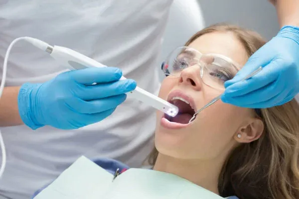
Visual Aid for Patient Education: Intraoral cameras allow patients to see a detailed and close-up view of their oral health. Dentists can capture real-time images of the inside of the mouth, such as teeth, gums, and other oral structures. By visually explaining dental conditions or treatment options, patients can better understand their oral health and make informed decisions about their dental care.
Early Detection of Dental Issues: Intraoral cameras can capture high-resolution images that reveal even the smallest dental problems that might go unnoticed during a routine examination. Early detection of issues like tooth decay, cracks, gum disease, and oral lesions allows for prompt intervention, preventing further complications and potentially reducing treatment costs.
Improved Communication: Intraoral cameras facilitate better communication between dentists and patients. By sharing images of specific areas of concern, dentists can effectively communicate the need for treatment and explain the recommended procedures. Patients can ask questions and actively participate in their treatment planning process, leading to a stronger dentist-patient relationship.
Accurate Treatment Planning: Intraoral cameras provide precise and detailed images, enabling dentists to accurately diagnose dental conditions and develop tailored treatment plans. With a clear visualization of the affected areas, dentists can plan and execute treatments with greater precision, leading to more successful outcomes.
Documentation and Tracking: Intraoral cameras capture images that can be stored in patients’ digital records for future reference. These images serve as valuable documentation of a patient’s oral health status over time, allowing dentists to track changes, monitor the effectiveness of treatments, and evaluate long-term oral health improvements.
Enhanced Patient Comfort: Intraoral cameras are small, handheld devices with a slim camera wand that can easily maneuver inside the mouth. They provide a non-invasive and comfortable experience for patients, minimizing discomfort and anxiety associated with traditional methods of examination.
Collaboration and Referrals: Intraoral camera images can be easily shared with dental specialists or other healthcare providers, facilitating collaboration and referrals when necessary. This seamless sharing of visual information helps in obtaining second opinions, coordinating multidisciplinary care, and ensuring comprehensive treatment for complex dental cases.
In summary, intraoral cameras are a valuable tool in dentistry that promotes patient engagement, early detection of dental issues, accurate treatment planning, and improved communication between dentists and patients. By utilizing intraoral cameras, dental practices can enhance the quality of care, provide a more personalized experience, and foster better oral health outcomes for their patients.
Panorex

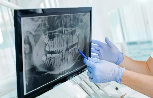
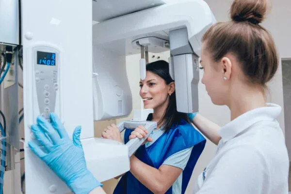
A Panorex, also known as a panoramic radiograph or panoramic X-ray, is a specialized dental imaging technique that provides a panoramic view of the entire oral and maxillofacial region in a single image. It captures a wide-angle view of the teeth, jaws, temporomandibular joints (TMJ), sinuses, and surrounding structures.
Here’s why a Panorex is important to have in a dental office:
Comprehensive View: A Panorex X-ray offers a comprehensive and detailed view of the entire oral cavity, allowing dentists to assess the overall oral health and detect various dental and skeletal abnormalities. It provides a broader perspective than traditional intraoral X-rays, enabling dentists to identify conditions that may not be visible with other imaging techniques.
Diagnosis of Dental Conditions: Panorex X-rays are instrumental in diagnosing a range of dental conditions and diseases. Dentists can detect issues such as impacted teeth, dental caries (cavities), periodontal disease, bone loss, cysts, tumors, jaw fractures, and developmental abnormalities. This comprehensive imaging helps in formulating accurate diagnoses and developing appropriate treatment plans.
Treatment Planning: The panoramic view obtained from a Panorex X-ray aids in treatment planning for various dental procedures. Dentists can assess the relationship between teeth, evaluate the bone structure, determine the presence of adequate bone for dental implant placement, and plan orthodontic treatment. This information ensures that treatment is tailored to the patient’s specific needs and optimizes treatment outcomes.
Identification of Impacted Teeth: Panorex X-rays are particularly useful in identifying impacted teeth, which are teeth that fail to erupt into their proper position. Common examples include impacted wisdom teeth (third molars) or canines. Accurate identification of impacted teeth helps dentists determine the need for extraction or orthodontic intervention to prevent complications and maintain oral health.
TMJ Evaluation: Panorex X-rays provide valuable information about the temporomandibular joints (TMJ) and their alignment. TMJ disorders, such as joint degeneration or misalignment, can be identified, leading to appropriate treatment planning or referral to a TMJ specialist.
Patient Education: Panorex X-rays serve as visual aids for patient education. Dentists can show patients their own X-ray images, explaining the specific dental conditions or treatment options visually. This helps patients understand their oral health status, the need for treatment, and the potential benefits of different procedures, enhancing their involvement in the decision-making process.
Referrals and Collaboration: Panorex X-rays are valuable diagnostic tools that can be shared with other dental specialists or healthcare professionals, facilitating referrals and collaborative care. When necessary, these images can be easily sent to oral surgeons, General Dentists, periodontists, or other specialists, ensuring comprehensive treatment and coordinated care.
In summary, a Panorex is an important imaging tool in dentistry due to its ability to provide a comprehensive view of the oral and maxillofacial region. It aids in diagnosis, treatment planning, identification of dental conditions, patient education, and collaboration with other healthcare providers. By having a Panorex machine in a dental office, dentists can provide more accurate diagnoses, tailored treatment plans, and improved patient care.
iTero scanner

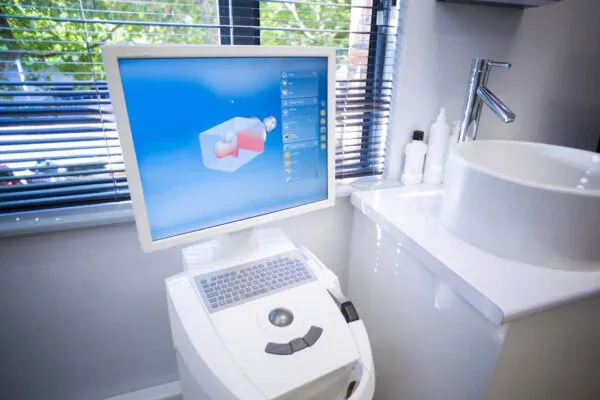
The iTero scanner is an intraoral digital scanner used in dentistry to create highly accurate 3D digital impressions of a patient’s teeth and oral structures. It offers several benefits for patients, enhancing their dental experience in the following ways:
Comfortable and Non-invasive: The iTero scanner replaces the need for traditional impression materials like alginate or putty. Instead, it uses a handheld wand to capture digital impressions of the teeth. This eliminates the discomfort and gag reflex often associated with traditional impressions, providing a more comfortable experience for patients.
Quick and Efficient: The iTero scanner captures digital impressions rapidly, significantly reducing the time required for the impression process. It eliminates the need for patients to sit with impression material in their mouth for an extended period, making the overall dental visit more efficient and time-saving.
Enhanced Accuracy: The iTero scanner creates highly precise and detailed digital impressions of the teeth, capturing even the smallest anatomical features. This accuracy ensures that restorations, such as crowns, bridges, or aligners, fit properly and provide optimal results. It helps minimize the need for remakes or adjustments, saving time and improving treatment outcomes.
Improved Visualization and Patient Education: The digital impressions captured by the iTero scanner can be immediately viewed on a computer screen. Dentists can visually explain the patient’s dental conditions, treatment options, and potential outcomes using the 3D digital models. This visual aid enhances patient understanding, enabling them to make informed decisions about their dental care.
Customized Treatment Planning: The digital impressions obtained from the iTero scanner can be used to create virtual models of the patient’s teeth. Dentists can manipulate these models to simulate various treatment options and show patients the potential results before initiating any procedures. This personalized treatment planning approach helps patients visualize their treatment journey and actively participate in the decision-making process.
Seamless Integration with Digital Dentistry: The digital impressions from the iTero scanner can be easily integrated into various digital workflows. They can be shared electronically with dental laboratories or specialists, facilitating accurate communication and collaboration. This seamless integration streamlines the entire treatment process and ensures consistent and high-quality results.
Follow-up and Monitoring: The iTero scanner enables dentists to compare digital impressions taken at different points in time. This feature is particularly useful for tracking the progress of orthodontic treatment or monitoring the effectiveness of dental restorations. Patients can visually see the changes in their oral health over time, enhancing their motivation and engagement in their dental care.
In summary, the iTero scanner offers numerous benefits to patients, including enhanced comfort, improved accuracy, time efficiency, better visualization, personalized treatment planning, and seamless integration with digital dentistry. By utilizing this advanced technology, dental professionals can provide a more comfortable, efficient, and patient-centered dental experience, ultimately leading to better treatment outcomes and patient satisfaction.
